Bryan Ng
This is a summary of a paper published by researchers from the University of Victoria including ICORD researcher Dr. Stephanie Willerth.
Khadem Mohtaram N, Ko J, King, C, Sun L, Muller N, Byung-Guk Jun M,Willerth SM. 2014. Electrospun biomaterial scaffolds with varied topographies for neuronal differentiation of human-induced pluripotent stem cells. J Biomed Mater Res Part A2014:00A:000–000. Find the Original Article here.
How Can Biomaterial Scaffolds Help Stem Cells Grow?
The ability of stem cells to develop into different types of cells, more importantly in our case into neurons for different medical applications, is promising. However, there is a challenge in controlling the growth of pluripotent (Cells with the ability to develop into any type of cell – from skin cells to neurons and everything in between) cells into 3D structures as one would find in a healthy body.
First, let’s have a very brief overview of two types of pluripotent stem cells:
- Embryonic Stem Cells: cells from human fertilized eggs that are capable of dividing and specializing into any cell type – controversial because they derive from a fertilized human egg
- Induced pluripotent stem cells: (iPSCs) created from non-reproductive human cells and are reprogrammed to act like Embryonic Stem Cells
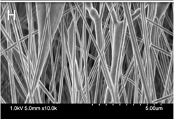
It is known that chemicals can guide the growth of stem cells; however, an overlooked but important factor is the physical environment that can also guide stem cell growth and specialization. The biomaterial technology used in this experiment, electrospun fibres (learn more about electrospun fibres here and in a previous blog entry) are used to create a scaffold structure (a sort of skeleton that helps create structure) and can be modified so that both chemical and physical factors are available to help guide the growth and specialization of stem cells in a 3D environment. This new study uses a method of creating microfibres called melt electrospinning that involves melting a polymer into a fluid that is then shaped into a microfiber. This method does not use toxic solutions to generate these fibres. These microfibers were then coated with a layer of nanofibers produced using solution electrospinning (a method of electrospinning that requires toxic solutions to create microfibers) to control the physical surface of the scaffold and create consistent products which simulate a 3D environment. The surface of the support structure produced is highly controllable, making it ideal for clinical applications.
What Does This Study Cover?
This study explores how the different physical surfaces of the electrospun fibre scaffold influences the amount of cell growth and level of specialization of stem cells into neurons.
The researchers focused on the fibre thickness in the scaffold structures and engineer two new fibre layouts, biaxial and bimodal aligned (defined below), to study the effects of physical cues on human iPSCs. The stem cells were cultured within the scaffold structures in order to see the varying effects physical cues around the cells had on the specialization and growth into neurons.
How Did The Researchers Conduct The Study And What Were The Results
Two different sized microfibres that made up different scaffolds were created using melt electrospinning:
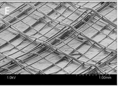
- Melt electrospinning was used to create the biaxial aligned scaffold. Biaxial means of two

Low magnification image of biaxial aligned scaffolds
axes; this scaffold has microfibres running vertically, as well as microfibres running horizontally, creating a criss-crossed surface
Results
- The microfibres that are thinner allow for better growth, spreading, and cell survival than thicker microfibres.
- Biaxial and bimodal scaffolds can support growth and specialization of stem cells into neurons.
- Biaxial & smaller diameter microfibres showed growth of more mature neurons.
- Overall, we find that fibre layout and size of microfibres influenced the differentiation of stem cells into neurons and direction of growth of neurons.
Things to consider
- Further studies are required to quantify the presence of all the different types of neural cells that differentiate on the scaffold.
- Melt electrospinning can consistently reproduce identical scaffold microfibres, making it a useful technique for bioengineering and clinical application.
- These scaffolds can hold molecules that promote the growth and specialization of stem cells into neurons.
What does this mean for people living with SCI?
The development of scaffolds that can guide stem cells with both physical and chemical cues means that we may soon be able to successfully grow neurons in a 3-dimensional structure as one would see in the human body. The growth of tissue/neurons in a 3-D environment may allow for cell/tissue repair or replacement with fewer complications for individuals living with SCI.


