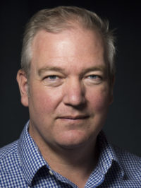
Principal Investigator
B.Eng. [Mechanical Engineering] (McGill University), Ph.D. [Engineering Science] (University of Oxford), Post-Doctoral Fellowship [Orthopaedic Biomechanics] (John Hopkins University), Post-Doctoral Fellowship (Harvard University)
Professor, Department of Orthopaedics, Faculty of Medicine, University of British Columbia
Associate Member, Department of Mechanical Engineering, Faculty of Applied Science, University of British Columbia
Research Interests
Biomechanics; Hip injury; Joints; MRI; OsteoarthritisDr. Wilson investigates the links between joint mechanics, clinical symptoms, and the success of treatment procedures. His research makes connections between biomechanics and problems like osteoarthritis and chronic pain. In his research, Dr. Wilson uses many medical imaging procedures, such as magnetic resonance imaging (MRI), computed tomography (CT), fluoroscopy, and ultrasound. He also works to develop new, more accurate methods for measuring the biomechanics of the spine, hip, and knee based on these imaging techniques.
Dr. Wilson is a Principal Investigator at ICORD, in addition to being a Professor in the Department of Orthopaedics at the University of British Columbia. He is also an Associate Member of the Department of Mechanical Engineering at the University of British Columbia. He obtained his B.Eng. in mechanical engineering at McGill University and his Ph.D. in engineering science at the University of Oxford. He also completed Post-Doctoral Fellowships at John Hopkins University and Harvard University.
Dr. Wilson’s work translates to functional improvements after SCI by improving surgical treatments for diseases and injury. Having a better understanding of the causes of these conditions allow researchers to outline risk factors and therefore provide more effective prevention and treatment strategies.
His favourite aspect of working with ICORD is being able to interact with trainees from all aspects and disciplines. Not to discount his colleagues, he assures us. He values their interaction too!
Dr. Wilson chose the University of Oxford to complete his Ph.D. partly to join their competitive rowing team. While he was part of the team he began to notice the biomechanical injuries and joint problems which resulted from the demanding process of rowing. Although Dr. Wilson already knew he was interested in engineering, this experience with rowing drew his attention directly toward biomechanical engineering and eventually led to his specialization in joint and bone.
Recent Collaborations:
Dr. Wilson recently collaborated with Dr. Pollard (University of Oxford) on hip mechanics and hip problems.
He has also worked with University of Sydney researchers on the mechanics of knee braces and how those braces change the functioning of the joint.
With Boston University and Harvard University scholars, Dr. Wilson used novel contrast agents to image cartilage.
Major Findings:
Dr. Wilson used an MRI technique to study hips with abnormalities. He found that hip deformities are associated with early arthritis and also established the usefulness of the imaging technique in assessing such problems.
He has also used MRI to assess the effect of knee braces on patients with osteoarthritis. He found that although bracing made changes in the motion of the joint, these changes were limited and did not reduce the pain experienced by the subjects.
With Dr. Bonnie Sawatzky, Dr. Wilson helped develop a new technique to detect bone density changes associated with arthritis.
For more of his major findings, please see the selected publications below, as well as his recent publications listed at the bottom of this page:
- Ligaments and articular contact guide passive knee flexion
- Effect of augmentation on the mechanics of vertebral wedge fractures
- Magnetic resonance imaging for in vivo assessment of three-dimensional patellar tracking
- Determinants of patellar tracking in total knee arthroplasty
- Computed tomography topographic mapping of subchondral density (CT-TOMASD) in osteoarthritic and normal knees: methodological development and preliminary findings
Techniques employed in the lab:
- Biplanar fluoroscopy
- Contrast-enhanced imaging
- In vivo studies and ex vivo experiments
- MRI
- Open MRI
- Quatitative MRI (qMRI)
Affiliations with organizations and societies:
- Centre for Hip Health and Mobility (CHHM)
Awards
Some of Dr. Wilson’s recent major awards and accomplishments include:
- Highly Commended Paper in the category of Measurement Science & Technology (Journal of the Institutes of Physics, 2009)
- Outstanding Trainee Presentation (International Society for Hip Arthroscopy Meeting, 2009)
- Outstanding Paper Award in the category of Measurement Science & Technology (Journal of the Institutes of Physics, 2008)
- Excellence in Research Award (American Orthopaedic Society for Sports Medicine, 2003)
Trainee Awards
| Year | Name | Award |
|---|---|---|
| 2021 | Emily Sullivan | CGSM Scholarship, NSERC |
| Masters Students | Ph.D Students | Research Staff |
|---|---|---|
| Emily Sullivan* | Luke Johnston | Amy Phillips |
| Edward Hoptioncann | Carly Jones | Honglin Zhang |
*has graduated in the past year
Current Opportunities in the Lab
There are currently no openings in Dr. Wilson’s lab. Please contact Dr. Wilson with inquiries.
Recent publications
- Whittaker, JL et al.. 2025. Digital education and exercise therapy versus minimal intervention for young people at high risk of early onset knee osteoarthritis after ACL reconstruction: a study protocol for the Stop OsteoARthritis (SOAR) randomized controlled trial.. Trials. doi: 10.1186/s13063-025-08896-6.
- Johnson, LG et al.. 2025. Early age-related changes to articular cartilage T1ρ in hips with Legg-Calvé-Perthes disease deformity.. Osteoarthr Cartil Open. doi: 10.1016/j.ocarto.2025.100589.
- Johnson, LG et al.. 2025. Anterior Hip Clearance in Residual Legg-Calvé-Perthes Disease.. J Pediatr Orthop. doi: 10.1097/BPO.0000000000002949.
- Sullivan, ES et al.. 2025. Relationship between magnetization transfer ratio and axial compressive strain in tibiofemoral articular cartilage.. J Mech Behav Biomed Mater. doi: 10.1016/j.jmbbm.2025.106937.
- Johnson, LG et al.. 2025. A framework for three-dimensional statistical shape modeling of the proximal femur in Legg-Calvé-Perthes disease.. Int J Comput Assist Radiol Surg. doi: 10.1007/s11548-024-03272-2.

